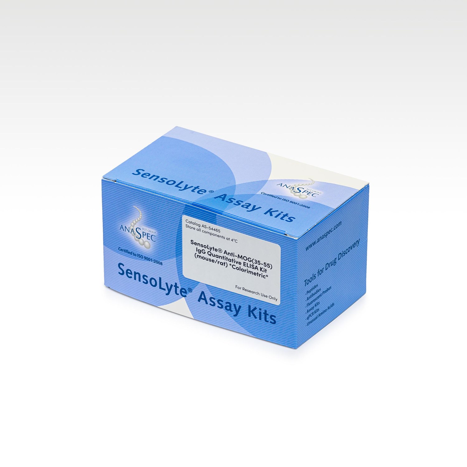
SensoLyte® Anti-MOG(35-55) IgG Quantitative ELISA Kit (mouse/rat) Colorimetric - 1 kit
- Cat.Number : AS-54465
- Manufacturer Ref. :
-
Availability :
In stock
- Shipping conditions : Ice fees will apply
Alternative choices

MOG peptide fragment (35-55) [Cat# AS-60130] induces autoantibody production and relapsing-remitting neurological disease causing extensive plaque-like demyelination. Autoantibody response to MOG (35-55) is present in multiple sclerosis (MS) patients and MOG (35-55)-induced experimental autoimmune encephalomyelitis (EAE) mice.
The SensoLyte® Anti-MOG (35-55) IgG Quantitative ELISA Kit (mouse/rat) provides a convenient, quantitative assay for detecting anti-MOG (35-55) IgG. This colorimetric assay, useful for determining the amount of anti-MOG (35-55) IgG present, can help provide information on the role anti-MOG (35-55) IgG plays in the development of EAE, an in vivo animal model for MS pathogenesis. The assays are performed in a convenient 96-well (strips format).
Specifications
| Packaging | |
| Kits components |
|
|---|---|
| Chemistry | |
| UniProt number |
|
| Storage & stability | |
| Storage Conditions |
|
| Activity | |
| Application | |
| Biomarker Target | |
| Detection Method | |
| Detection Limit |
|
| Research Area | |
| Sub-category Research Area | |
| Usage |
|
Downloads
You may also be interested in the following product(s)
SensoLyte® Anti-Mouse MOG (1-125) IgG Quantitative ELISA Kit Colorimetric - 1 kit
SensoLyte® Anti-PLP (178-191) IgG Quantitative ELISA Kit (mouse) Colorimetric - 1 kit
Citations
Isolating and targeting a highly active, stochastic dendritic cell subpopulation for improved immune responses
Cell Rep . 2022 Nov 01 ; 41(5) 111563 | DOI : https://doi.org/10.1016/j.celrep.2022.111563
- P. Deak
- et al
Dysregulated follicular regulatory T cells and antibody responses exacerbate experimental autoimmune encephalomyelitis
J Neuroinflammat . 2021 Jan 19 ; 18 27 | DOI : https://doi.org/10.1186/s12974-021-02076-4
- L. Luo
- et al
Myelin Basic Protein Phospholipid Complexation Likely Competes with Deimination in Experimental Autoimmune Encephalomyelitis Mouse Model
ACS Omega . 2020 Jun 30 ; 5(25) 15454 | DOI : 10.1021/acsomega.0c01590
- A. Valdivia
- et al
Follicular regulatory T cells can be specific for the immunizing antigen and derive from naive T cells
Nature Commun . 2016 Jan 28 ; 7 10579 | DOI : https://doi.org/10.1038/ncomms10579
- M. Aloulou
- et al
High Affinity Binding of Conformationally Constrained Synthetic Oligomers To An Antigen-Specific Antibody: Discovery of a Diagnostically Useful Synthetic Ligand For Murine Type 1 Diabetes Autoantibodies
Bioorg Med Chem Lett. . 2015 Nov 01 ; 25(21) 4910 | DOI : 10.1016/j.bmcl.2015.05.046
- TM. Doran
- et al
Cerebrospinal fluid dendritic cells infiltrate the brain parenchyma and target the cervical lymph nodes under neuroinflammatory conditions
PLoS ONE . 2008 Oct 02 ; 3 e3321 | DOI : 10.1371/journal.pone.0003321
- E. Hatterer